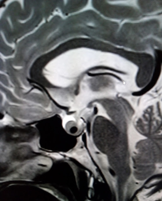
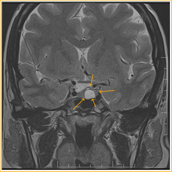
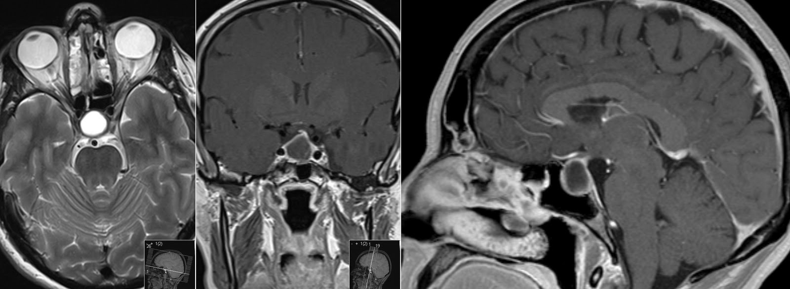
Rathke's Cleft Cyst Diagram
Mri and pathological features of rathke cleft cysts in the sellar region Rathke cyst cleft radiopaedia cystic intracranial tumors Rathke cyst cleft t1 sagittal radiology obtained weighted before brain mass various enhancement sellar extending marginated contrast depicts well into
Rathke's Cleft Cyst :MRI - Sumer's Radiology Blog
Figure 1 from suprasellar rathke cleft cysts: clinical presentation and Rathke's cleft cyst :mri Rathke zyste mri bild hypophyse kontrastmittel inselspital zysten neurochirurgie bern ausdehnung zeigt bereich
Rathke cleft cyst: practice essentials, radiography, computed tomography
Sagittal enhanced t 1 -weighted image of a rathke’s cleft cyst. theRathke cleft cyst Cyst cleft rathke mri pituitary pouch gland stalk radiology remnants compressionRathke's cleft cyst :mri.
Cleft rathke cyst mri radiologyRathke's cleft cyst Rathke's cleft cyst: mriRathke cyst cleft pituitary pouch anvekar frcr balaji dr sellar.

Surgical neurology international
Mri rathke cleft rcc intracystic cysts nodules cyst observed t2wi correspond pathological sellar etmSagittal mri of patient 20 years later noting return of the rathke's Dr balaji anvekar frcr: rathke’s cleft cystRathke's cleft cyst.
Cyst cleft rathke mri cysts brain healthRathke's cleft cyst Rathke cyst cleftCyst cleft rathke mri pituitary radiology.

Radiopaedia cyst rathke cleft radiology nodule intracystic
Cyst pituitary rathke cleft gland cysts mri radiology cystic sign lesion claw velum tumor symptoms cavum largeRathke's cleft cyst Rathke cleft cystRathke cleft suprasellar cysts figure treatment clinical outcomes presentation.
Rathke's cleft cyst-mriRathke-zysten – diagnose & therapie Cyst mri rathke sagittal notingRathke cleft cyst.

Cyst cleft rathke sagittal weighted enhanced enhancing
Rathke cyst cleft pituitary radiopaedia apoplexy size radiology gland version hemorrhagicCyst rathke cleft Cyst cleft rathke mri pituitary radiology pouch gland hyperprolactinemia compression stalk.
.


